
von
Anthocyanins represent a prominent class of water-soluble polyphenolic pigments responsible for the vivid red, purple, and blue coloration observed across a wide range of fruits and vegetables[1,2]. Within nutritional biochemistry and phytochemistry, the terms anthocyanin and anthocyanidin are frequently mixed, despite denoting distinct chemical structures with significantly different physical and physiological properties[3].
At the molecular level, the primary difference comes down to the presence or absence of attached sugar molecules, known as glycosidic linkages[4]. Understanding this structural architecture—specifically the central core and its side-chain modifications—is essential before we can examine how these compounds are broken down, absorbed, and metabolized in the human digestive tract.
1. The Fundamental Anthocyanin Core Structure

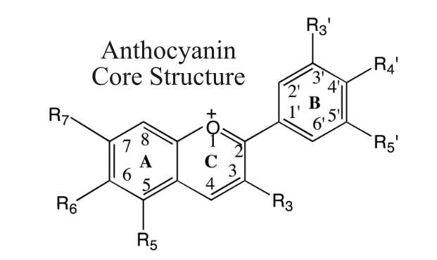
The framework of the Anthocyanin core is built around three distinct carbon rings—labeled A, B, and C[5]. Rings A and C are fused together on the left, while Ring B hangs off to the right like a side branch. What makes this central core particularly unique is the positive charge (+) sitting on the oxygen atom in Ring C[4,6]. This tiny electrical charge acts as the molecule’s color switch, allowing it to change shades when exposed to different environments (like shifting pH levels in plant sap or your stomach)[6].
The branches labeled with „R“ (R3, R5, R7 etc.) represent customizable attachment points. Nature attaches different chemical groups onto these specific positions to create different final molecules[1,4]. Substituting a simple hydroxyl group (-OH) for a sugar or methyl group changes everything from the compound’s final color (vibrant red versus deep violet) to how easily your body can absorb it[1,4].
2. Variations on the Core: The Six Primary Anthocyanidins
Before a sugar unit is attached to complete a full anthocyanin molecule, we are left with just the bare, sugar-free chemical core. In biochemistry, this central parent structure without any attached sugars is known as an aglycone (or anthocyanidin)[1,4]. Nature creates incredible color diversity right at this base level simply by substituting the functional groups attached to the core. While dozens of these sugar-free cores exist in nature, six primary aglycones account for roughly 90% of all natural plant pigmentation[1,4].
These six variants differ primarily by what occupies the R3′ and R5′ slots on Ring B, as well as the R3 slot on Ring C:
1. Pelargonidin

Absorbs light in a way that yields bright orange to brick-red hues. Sources: Strawberries, red radishes, and pelargonium flowers[1,4].
2. Cyanidin

Produces rich magenta, deep red, and reddish-purple tones. Sources: Blackberries, elderberries, red cabbage, and apples[1,4].
3. Delphinidin

Shifts light absorption toward deep blue, violet, and purple spectrums. Sources: Blueberries, Concord grapes, and eggplants[1,4].
4. Peonidin

Gives a bright purplish-red coloration. Sources: Peonies, cranberries, and red grapes[1,4].
5. Petunidin

Imparts deep purple to dark blue hues. Sources: Petunias, black beans, and blueberries[1,4].
6. Malvidin

Responsible for intense blue-violet and dark red shades; highly stable against oxidation. Sources: Red wine, blueberries, and dark grapes[1,4].
Looking across these six structures, a clear pattern emerges: the core scaffold remains identical, while small changes to the side groups on Ring B dramatically alter the pigment’s shade. However, in a living plant cell, these bare cores rarely exist on their own[1,2,4]. Without an attached sugar, free anthocyanidins are hydrophobic and chemically unstable in the neutral-to-acidic environment of plant sap[1,4]. Exposed to water and light, the central ring can rapidly degrade or lose its color[2,4].
To overcome this instability, plant enzymes perform a crucial chemical modification: glycosylation. By attaching a sugar molecule onto one of the open hydroxyl slots on the core scaffold, the plant transforms a reactive anthocyanidin into a highly stable, water-soluble anthocyanin[1,2,7].
3. Glycosylation: From Aglycone to Anthocyanin
The enzymatic process of attaching sugar units to the core aglycone scaffold is known as glycosylation[1,2]. This single structural change completely alters the physical behavior of the pigment—turning an unstable, water-insoluble core into a resilient, water-soluble molecule ready for storage in plant tissue[1,2].
Sugar Attachment and Numbering
While glucose is by far the most common sugar used by plants, others like galactose, rhamnose, arabinose, and xylose are also frequently utilized[1]. Nature varies both the location and the number of attached sugars to create hundreds of distinct anthocyanin molecules:
Monoglycosides (Single Sugar): The most fundamental and abundant setup in nature occurs when a single sugar molecule attaches to the C3 position on Ring C. Cyanidin-3-glucoside shown below is an example of a Monoglycoside[8].

Diglycosides (Two Sugars): Plants often attach two sugar units. This can happen in two distinct ways:
3,5-diglycosides: One sugar attaches to the C3 position on Ring C, and a second sugar attaches to the C5 position on Ring A [9].

3-disaccharides: Two sugars are linked directly to each other in a chain, which is then attached as a single unit to the C3 position (such as cyanidin-3-rutinoside)[10].

Fig 4. Cyanidin 3-O-rutinoside [10]
However, this same sugar attachment significantly hinders direct absorption in the human small intestine. [2] Because complex sugar-bound molecules cannot easily pass across the intestinal lining via simple diffusion, the anthocyanin remains largely intact as it travels through the upper digestive tract, allowing a major portion to reach the colon, where specialized gut microbes can finally cleave the sugar and break down the core structure.[2]
Triglycosides (Three Sugars): In more complex plant structures, three sugars can be incorporated—either attached across multiple positions (C3, C5, and C7)or built as longer disaccharide/trisaccharide chains at C3[10].
4. Acylation: Adding Organic Acids for Maximum Stability
Why Acylation Matters: Color and Heat Resistance
Acylation represents the highest tier of natural structural modification. Non-acylated anthocyanins (like those in strawberries or raspberries) degrade easily when exposed to heat, light, or pH changes during processing and cooking[1,2].
In contrast, heavily acylated anthocyanins—such as those found in red cabbage, purple sweet potatoes, and purple carrots [12]—are exceptionally stable. They preserve their vibrant purple and blue colors even during thermal processing and across a wider range of digestive pH conditions [2, 12].
Acylation is the process of attaching organic acid groups (acyl groups) directly onto the hydroxyl groups of the sugar molecules themselves—not directly to the flavylium core. [1,2] When an anthocyanin has one or more organic acids attached, it is called an acylated anthocyanin [1].
1. Aromatic Acyl Groups: These include hydroxycinnamic acids such as p-coumaric, caffeic, ferulic, and sinapic acids[1]. Because these organic acids contain their own aromatic ring structures, they fold over the central core like a molecular shield (a process called intramolecular copigmentation)[13]. This physical stacking protects the positively charged Ring C from water attack, preventing the molecule from losing its color or degrading.

Fig. 5 Chemical structure of Delphinidin-3-O-(6-p-coumaroyl)glucoside[12]
2. Aliphatic Acyl Groups: These include dicarboxylic acids such as malonic, acetic, oxalic, and succinic acids. While they lack aromatic rings, aliphatic acids alter the electrical charge and overall acidity of the molecule, enhancing its solubility and changing how it interacts with cellular membranes [14].

Fig 6. Chemical structure of cyanidin-3-(6“-O-malonylglucoside), an anthocyanin acylated with an aliphatic malonyl group [14].
5. Summary: The Structural Hierarchy
To bring this chemical overview together, nature builds these brilliant plant pigments using a three-tiered modular assembly line:
1. The Core Scaffold (Aglycone / Anthocyanidin): The three-ring core serves as the baseline framework. Small variations in side-chain groups on Ring B yield the six major aglycones (from the brick-red of Pelargonidin to the deep blue of Delphinidin).
2. Glycosylation (Anthocyanin): Attaching one or more sugar molecules (most commonly at position C3) converts the reactive, water-insoluble aglycone into a stable, water-soluble anthocyanin ready for plant storage.
3. Acylation (Acylated Anthocyanin): Decorating those attached sugars with aromatic or aliphatic organic acids adds an extra layer of protection, dramatically boosting color stability and resistance to heat and pH shifts.
6. Bibliography
1- He, J. et al. Anthocyanins: natural colorants with health-promoting properties. Annu. Rev. Food Sci. Technol. 2010, 1, 163–187.
2- Tena, N. et al. State of the Art of Anthocyanins: Antioxidant Activity, Sources, Bioavailability, and Therapeutic Effect in Human Health. Antioxidants 2020, 9, 451.
3- Sadowska-Bartosz, I. et al. Antioxidant Activity of Anthocyanins and Anthocyanidins: A Critical Review. Int. J. Mol. Sci. 2024, 25, 12001.
4- Khoo, H.E. et al. Anthocyanidins and anthocyanins: colored pigments as food, pharmaceutical ingredients, and the potential health benefits. Food Nutr. Res. 2017, 61, 1361779.
5- Alshamar, H.A. et al. Anthocyanins from a single botanical source can be used as a replacement for hemalum and eosin. Biotech. Histochem. 2021, 96, 570–578.
6- Basílio, N. et al. Chemistry and Photochemistry of Anthocyanins and Related Compounds: A Thermodynamic and Kinetic Approach. Molecules 2016, 21, 1502.
7- Li, Z. et al. Assessing Anthocyanin Biosynthesis in Solanaceae as a Model Pathway for Secondary Metabolism. Genes 2019, 10, 559.
8- Olivas-Aguirre, F.J. et al. Cyanidin-3-O-glucoside: Physical-Chemistry, Foodomics and Health Effects. Molecules 2016, 21, 1264.
9- He, F. et al. Anthocyanins and Their Variation in Red Wines I. Monomeric Anthocyanins and Their Color Expression. Molecules 2012, 17, 1571–1601.
10- Horiuchi, R. et al. Identification of the Biosynthetic Pathway for Anthocyanin Triglucoside, the Precursor of Polyacylated Anthocyanin, in Red Cabbage. J. Agric. Food Chem. 2020, 68, 9750–9758.
11- Oancea, S. et al. A Review of the Current Knowledge of Thermal Stability of Anthocyanins and Approaches to Their Stabilization to Heat. Antioxidants 2021, 10, 1337.
12- Costa-Pérez, A. et al. The (Poly)phenolic Profile of Separate Winery By-Products Reveals Potential Antioxidant Synergies. Molecules 2023, 28, 2081.
13- Ricci, A. et al. The Role of Copigmentation in Colour Attributes and Their Evolution in Model Wine: A Thermodynamic and Colorimetric Study. Foods 2025, 14, 2467.
14- Alappat, B. et al. Anthocyanin Pigments: Beyond Aesthetics. Molecules 2020, 25, 5500.